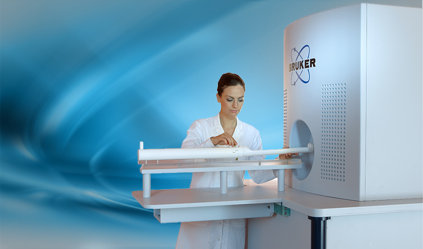
“Czech Republic Gets 3rd in the world Magnetic Particle Imaging Scanner”
For the first time, a commercial Magnetic Particle Imaging (MPI) scanner is up and running outside the country of Germany, where MPI was developed. The MPI is the centerpiece of the new multimodal Center for Advanced Preclinical Imaging at the First Faculty of Medicine, Charles University, Prague, Czech Republic. It is only the third commercial MPI scanner in the world.

Guests attend the opening of the Center for Advanced Preclinical Imaging at the First Faculty of Medicine, Charles University, Prague, Czech Republic
Magnetic Particle Imaging is a fundamentally new way of seeing in vivo functional activity. It complements other preclinical imaging systems to study diagnostic techniques and treatment approaches that have a high possibility of translating to a clinical setting. The hope is that the MPI technology for medical and industrial research will ultimately move from the preclinical setting and be used in patient care. Research areas include cardiovascular disease, cancer and stem cell therapies.
Magnetic Particle Imaging is a new tomographic imaging method that rapidly captures in vivo 3D images at millisecond intervals using magnetic tracers. The tracer is a super paramagnetic iron-oxide nanoparticle. By injecting the tracers into the bloodstream or attaching them to cells or drugs of interest, it becomes possible to follow activity and movement in real time. The MPI system, developed by Bruker BioSpin and Royal Philips Electronics, allows rapid and accurate tracking of super paramagnetic particles on a magnetic resonance imaging (MRI) background.
Along with the new Magnetic Particle Imaging scanner, the Center for Advanced Preclinical Imaging (CAPI) houses other innovative preclinical imaging solutions from Bruker to enhance the study of small animals. The center has Micro-CT (Computed Tomography), PET (Positron Emission Tomography), SPECT (Single-Photon Emission Computed Tomography), MRI (Magnetic Resonance Imager), Optical Imager (fluorescence, luminescence on X-ray background.) The center will acquire a high-frequency ultrasound (US) with photoacustic mode, a confocal endoscope and other instruments by the end of 2017.
![]()
Having the equipment in one location means researchers can combine information and views from a number of systems to create more complete and more informative images than previously possible.

CAPI houses a wide variety of preclinical imaging instrumentation
Research areas
MPI can produce real-time, high-resolution images that capture cardiovascular activity in a mouse. Such excellent temporal resolution (precise image information in real time) is unique to MPI, and may prove useful in understanding complex cardiovascular functions, measuring ejection fraction and locating specific vessels where blood flow is weak.
Right now, cardiac x-ray angiography is the standard way to assess coronary obstructions. While the technique produces real-time, high-resolution images with good contrast, the 50-year-old technology comes with risks inherent in radiation and contrast agents, which may cause kidney damage or allergic reactions. By comparison, angiography by magnetic particle imaging could provide excellent images more safely.
Stem cells hold great promise for treating a variety of illnesses, such as vascular disease and cancer, and injuries, such as burns and spinal damage. But to make the most of that promise, researchers need to visualize the stem cells, to see that they move to the targeted location, remain alive and provide benefits. The ability to mark individual cells with magnetic particles and track them in vivo makes Magnetic Particle Imaging ideally suited to the job.
MPI may help study muscarinic receptors in mice that have deficiencies associated with central nervous system disorders, such as Alzheimer’s disease. It also will help track photosensitizing agents as they travel to cancer cells, ensuring the efficacy of photo chemotherapy methods.
In the future
The results from the preclinical MPI research in Germany, and starting this month from Prague, will improve MPI technology and provide vital information on how to bring the benefits of MPI to patients in the future. The unique capabilities of MPI might one day improve diagnosis and treatment of people with heart and blood vessel diseases, cancer, burns and a host of other challenges.
For more information
Panagiotopoulos N, Duschka RL, Ahlborg M, et al. Magnetic particle imaging: current developments and future directions. International Journal of Nanomedicine. 2015;10:3097-3114. doi:10.2147/IJN.S70488.
Preclinical Magnetic Particle Imaging: Interview with Jeff W. M. Bulte, M.S., Ph.D., Johns Hopkins Medicine
- Professor in the Johns Hopkins Departments of Radiology, Oncology, Biomedical Engineering and Chemical & Biomolecular Engineering.
- Director of Cellular Imaging at the Johns Hopkins Institute for Cell Engineering.
