A Free Preclinical Imaging Educational Webinar
This webinar was recorded on May 12, 2015. Click to watch the recording!
Seminar Overview
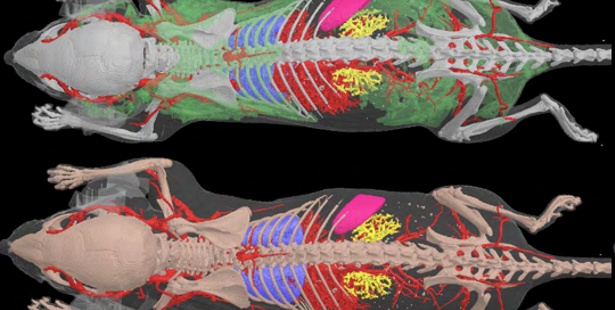
The use of X rays in preclinical applications is historically linked to imaging bone, where it has become the gold standard. X rays are attenuated more by calcified tissue compared to soft tissue. However, in recent years, the range of applications has greatly increased through the development of preclinical contrast agents. This has made it possible to apply the microCT technology in soft-tissue applications such as lung imaging, cardiovascular applications and oncology.
In vivo imaging of mice and rats at high resolution with low X rays dose allows for longitudinal follow up of disease progression. In addition the researchers can monitor drug treatments at multiple time points, giving them a full understanding of the kinetics.
As microCT imaging is a non-destructive technology, tissues can also be scanned ex vivo with sub-micron pixel size generating images similar to histological sections, but adding the third dimension. Researchers no longer have to choose which cross-section to select but have access to the full three dimensional volume. This 3D information is crucial when screening tissues for e.g. tumors, or for looking for diseases which are non-uniformly spread within an organ.
Who should attend
Any Lab Managers, Scientists, Researchers, Technicians, Students working in Academia, Hospitals or Institutions currently interested in the expanding field of microCT for preclinical Applications.
- Research group leaders
- Laboratory managers
- Biomedical Scientists
- microCT Specialist
- Oncology Scientists
- Bone Researchers
- Cardiology Scientist
- Lung Scientist
Presenters
Jeroen Hostens, PhD
Applications Manager, Bruker microCT
Kjell Laperre, PhD
Applications Scientist, Bruker microCT
