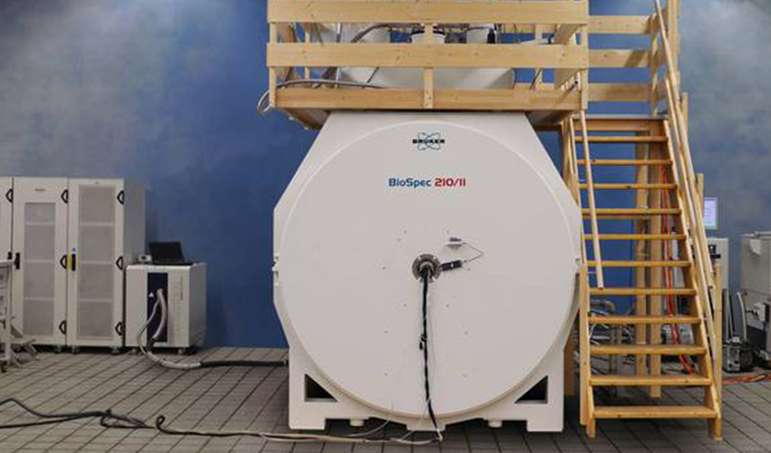
MRI systems are now commonly used in hospitals and medical centers around the world for medical diagnostic imaging. Despite this ‘applied’ market being long established and well served by a number of manufacturers, ‘high field’ imaging is still only an emerging area in medical and veterinary research. Following the development of the world’s first 21 T horizontal magnet, Tim Wokrina, Product Manager for Bruker BioSpin, discusses the trend towards ultra high field imaging, examining the potential benefits and challenges of this cutting-edge technology.
Magnetic resonance imaging technologies have developed rapidly over the last decade. These advances have led to MRI systems becoming increasingly affordable, with both large bore 1.5 T and higher resolution 3.0 T platforms now a common sight in medical imaging suites around the world. Beyond these ‘routine’ imaging systems, there is a growing trend towards the use of ‘high field’ MRI technologies for scientific research. This exciting approach has the potential to further our understanding of a wide range of biological processes and diseases, providing real-time in vivo imaging to elucidate changes at the cellular level.
Pushing the boundaries
Preclinical and molecular MRI is a fast growing market segment in medical and veterinary research. Combining ultra high field (UHF) magnets – field strengths of 11.7 T or greater – with state-of-the-art low temperature RF coils and preamplifiers, these systems offer exceptional spatial resolution and contrast for in vivo imaging. The availability of imaging platforms such as the 15.2 T BioSpec® 152/11 USR/R (Bruker) as ‘off-the-shelf’ products means that facilities around the world are now regularly reporting discoveries that push the boundaries of what can be achieved using MRI in a research or preclinical setting.
However, as anyone routinely working with MRI systems will tell you, a new field strength brings its own challenges as well as advantages. For example, the difference between 1.5 T and 3.0 T systems, even with commercially available diagnostic imaging platforms, is more than simply an increase in resolution. The changing magnetic field strength affects the signaltonoise ratio, contrast and appearance of artefacts. It is therefore very important to understand any newly developed system from a technical perspective before beginning to explore its application-oriented capabilities. To simplify this characterization, Bruker’s UHF superconducting magnets are constructed using a modular design that is equally suited to imaging or mass spectrometry applications, allowing the Bruker team to validate each new unit using its BioSpec MRI platform before shipping to the customer.
Based on state-of-the-art MRI CryoProbe™ technology, the BioSpec series is designed for the emerging markets of preclinical and molecular MR imaging, and combines the latest HD electronic architecture with digital filtering, quadrature demodulation and RF amplitude stabilization to offer superior resolution and image quality. This modular set-up is ideally suited to the rapid integration and validation of new UHF magnets, providing a robust platform for instrument and application development.
A new benchmark
The development of the world’s highest field horizontal magnet – a 21 T, 11 cm bore superconducting magnet – at Bruker’s Karlsruhe R&D and production facility is a landmark in this exciting area of research, and provided an exciting opportunity to explore the unparalleled imaging capabilities of this field strength.
Designed to be the heart of a new FT-ICR (Fourier transform ion cyclotron resonance) mass spectrometer in the National High Magnetic Field Laboratory (NHMFL) at Florida State University, this record-breaking magnet was coupled with a custom-built 900 MHz MRI CryoProbe to create near-cellular resolution in vivo images of the mouse brain for the first time. Using this set-up, the Bruker team was able to achieve a ‘slice’ thickness of just 26 µm, virtually eliminating the need for traditional histological approaches in many applications.
MR histology – the future of preclinical research?
From the perspective of preclinical MRI, achieving true cellular resolution is the ultimate goal, allowing in vivo drug studies to be performed longitudinally in a single organism. Traditional histological approaches – euthanizing the test subject, sectioning, mounting and staining material, then observing the results under a microscope – are both time consuming and extremely expensive, as well as raising ethical concerns. The ability to avoid this – as well as to use each animal as its own control by establishing a baseline prior to drug administration – has huge potential advantages for the industry, and the preliminary findings of the 21 T magnet validation study highlight the potential of this approach for accelerating drug development. We are only just beginning to explore the potential of high field MRI, but the future is very exciting.
