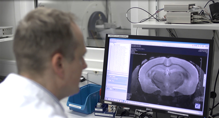
Preclinical imaging is an essential tool for observing changes in organs, tissues, cells and even molecules in animal models, often in response to physiological and environmental stimuli.
Recent technological advances in preclinical MRI imaging have increased the quality and extent of research that can be carried out in this field. The technique offers high spatial resolution imaging that can be applied to a broad range of areas, from very basic research, right up to applied science.
Located at The University of Freiburg in Germany, is the Advanced Molecular Imaging Research Center (AMIR), where scientists focus on multimodal imaging for preclinical and translational research and the development of novel techniques that can be applied in small animal imaging.
Heading the center is Priv. Doz. Dr. Dominik von Elverfeldt, who recently talked about some of the current neurology projects underway at the center and what he finds particularly exciting about the advancing field of preclinical MRI imaging:
“The big advantage of MRI is its diversity in contrasts. You can basically tailor your sequence towards the task you have… whether you want to measure diffusion or perfusion; whether you want to know which brain areas work during a certain task; or whether you want to know how blood flows in the arteries.”
Lately, AMIR scientists have made promising new developments in the fields of resting state functional MRI (R-fMRI) and diffusion tensor imaging (DTI). Functional MRI provides information about brain function. The difference between traditional fMRI and R-fMRI is that fMRI involves the study of brain activity in response to a particular stimulus. R-fMRI, on the other hand, is concerned with observing brain activity in the absence of a stimulus, recording that activity over time and then gathering information about which brain areas communicate with one another.
DTI is a recently developed MRI-based neuroimaging technique that can measure axonal organization in tissues of the nervous system. “DTI allows you to measure the spatial resolution of the diffusivity of water, especially within the brain. With this, you are able to calculate where your nerve fibers run, so you’re able to get a map of the architecture of the brain,” explains Elverfeldt.
Information about the structural and functional networks within the brain and how they interact enables the team to investigate changes that occur in conditions such as multiple sclerosis, depression and addiction. It also helps them to understand the potential treatment options for such diseases and disorders.
When a person develops an addiction, functional changes occur in the brain that eventually lead to structural changes. At the moment, Elverfeldt and team are looking for those changes in an animal model of alcohol addiction.
In another project, the researchers are using DTI to detect the loss of myelin that occurs in multiple sclerosis and are currently exploring the use of an artificial hormone that they expect will induce remyelination. If this study produces positive results, a clinical trial should directly follow.
The team has also been working on defining brain areas that are affected in depression because targeted stimulation of those areas could dramatically improve the lives of depressed individuals.
Understanding the networks in the brain and how they interact would also be useful to neurosurgeons, who could then ensure that critical brain areas are spared during surgery. This information may also reduce the time required to perform such procedures.
Elverfeldt described what he feels has been the most outstanding technological advancement to help the team over recent years, which was the development of a cryogenically-cooled RF coil. One of the main factors that effective imaging depends upon is a high signal-to-noise ratio (SNR; the ratio of the signal strength of the image to background noise). When component RF coil sensitivities are inadequately characterized or not distinct enough from one another, this can result in a degraded SNR, but cooling the coil improves the SNR by reducing resistive losses in it.
“This reduces the electronic noise drastically and thus leads to a gain in signal-to-noise ratio between a factor of two and three. Only that higher sensitivity allows us to acquire high quality data in a shorter time and only that yields the state-of-the-art resting state and DTI data,” says Elverfeldt.
Elverfeldt expects the future of preclinical MRI imaging to be bright, not only because of the potential for investigating disease and finding and monitoring treatments, but also in terms of how it could improve personalized medicine.
In the field of oncology, for example, tumors differ between patients. The interactions between tumors and patients also vary. If it was possible to take a specimen from a tumor and test different treatments on it, therapies could be tailored on an individual basis, which would significantly improve treatment success rates.
For more information about Elverfeldt’s work and the projects underway at AMIR, please visit the following link: https://www.uniklinik-freiburg.de/mr-en/research-groups/amir.html
