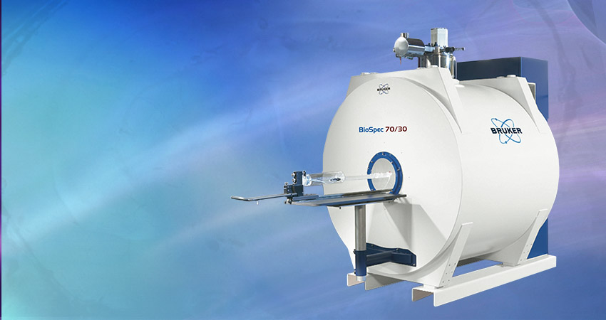
The Weizmann Institute completes installation of system following start of EU grant funding for multicolor imaging with MRI.
The Weizmann Institute of Science in Rehovot, Israel, recently announced the installation of the Biospec® 15.2 Tesla Avance III HD preclinical ultra-high field magnetic resonance imaging (MRI) from Bruker (NASDAQ: BRKR). The new instrument is installed in the Daniel Wolf building of the Institute’s Chemical Research Support Department, and will be used by leading MRI and biology research groups including among others those of Dr. Amnon Bar-Shir, Dr. Assaf Tal, Prof. Lucio Frydman and Prof. Michal Neeman, to open new frontiers in molecular and microscopic imaging.
The new 15.2 Tesla MRI is the latest MRI instrument from Bruker to be installed on site in Israel, and will operate alongside the Bruker BioSpec 4.7 Tesla and Bruker BioSpec 9.4 Tesla horizontal MRIs, and a Bruker 400 MHz wide-bore microimaging spectrometer. The cryoprobe-equipped 15.2T instrument will enable new forms of multiplexed imaging not previously available to the Institute, with the aim to develop, optimize and implement genetically engineered reporter systems for MRI with artificial “multicolor” characteristics. The instrument will also deliver superior spectral resolution for functional brain studies, the capability of studying low-gamma insensitive nuclei, and serve as imaging platform for an accompanying dissolution DNP hyperpolarizer. The first high-quality in vivo images have already been obtained in October 2017.
The Weizmann Institute of Science is one of the world’s leading multidisciplinary basic research institutions in the natural and exact sciences. It is a world-renowned center for magnetic resonance; its tradition of nuclear and electron magnetic resonance theory and experiment date back to S. Meiboom. Prof. Frydman is a leader in the fields of NMR and MRI, whose research focuses on developing new methodologies for accelerating the acquisition, improving the sensitivity and enhancing the resolution of both spectra and images.The Neeman lab has a track-record in multi-modal biological imaging, leveraging the MRI’s advantages together with customized contrast agents and with other optical modalities in order to monitor in vivo biological variables non-invasively. These include tracking the progression and regulation of angiogenesis and cell migrations in cancers and pregnancies, providing insight of biological and translational relevance. The Bar-Shir lab aims to develop new biosensors and tracers for MRI applications by using synthetic chemistry, nanotecnology and molecular biology approaches. Such biosensors will help scientists around the world to study unrevealed biological events longitudinally, non-invasively in deep tissues of a live subject. The Tal lab is focused on providing new MRS tools and insights that, by complementing traditional functional MRI pictures, will enable the use of spectroscopic markers for understanding brain metabolism in healthy and diseased states.
By bringing together a group of talented scientists working in all areas of MRI – from physical and image processing aspects to chemical development and applications in varied biological contexts – and endowing them with the most advanced imaging instrumentation, the Weizmann Institute is displaying scientific leadership and vision. Frydman says: “The infrastructure that is being created here will allow us, in combination with novel ultrafast MRI and MRS methods, progress in nuclear hyperpolarization, the development of new contrast agents and CEST sensors, and advanced biological techniques, to monitor a wide range of biologically relevant processes. Furthermore, the ultrahigh fields in question will allow us to measure a unique array of metabolic variables, while linking them with morphological tissue information and with dynamic information about perfusion and diffusion. This is truly unique information that will surely revolutionize our understanding about cellular mechanisms, and the function of healthy organs, and will have important consequences in terms of elucidating the onset and progression of diseases.”
Amnon Bar-Shir, Ph.D., Department of Organic Chemistry, Weizmann Institute of Science stated: “We feel this new installation will be a real game changer for our facility. The new MRI will give us superior spectral resolution and boost the signal-to-noise ratio for higher spatial resolution and faster measurements. The new instrument will facilitate the research of our world-class team and will reinforce Weizmann’s position as one of the premier centers in the world for MR research.” Assaf Tal, PhD, of the Insitute’s Chemical and Biological Physics Department adds: “The added sensitivity boost afforded by the ultrahigh field is essential for obtaining accurate spectroscopic information about brain metabolism, specifically observing important neurotransmitters such as glutamate and GABA at anatomically meaningful scales and during functionally activation. This is a major step forward in our ability to understand basic brain physiology.”
Wulf-Ingo Jung, Ph.D., the President of the Bruker BioSpin Preclinical Imaging division, added: “We are extremely pleased to partner with the Weizmann Institute and provide the enabling MRI research tools they need to advance the scientific understanding of the biomolecular processes of many diseases, as well as the understanding of advanced bio-materials that will impact many emerging diagnostic and therapeutic approaches.”
The BioSpec® product series is designed for the emerging market of preclinical and molecular MR imaging and MRI research. State-of-the-art MRI CryoProbe™ technology combined with ultra-high field USR magnets deliver high spatial resolution in vivo at highest stability, thus enabling customers to come closer to the molecular and cellular level research they desire. Due to the largest preclinical portfolio of sequences and in vivo optimized measurement protocols, virtually any small animal MR imaging application in life science, biomedical and preclinical research can be conducted.
