“Biomedical research relies on the use of animal models to understand the pathogenesis of human disease at the cellular and molecular levels, and to provide systems for developing and testing new therapies. Mammalian models, such as the mouse, have been pre-eminent in modeling human diseases, primarily because of the striking homology between mammalian genomes and the many similarities in the biology of mice and human beings, spanning from anatomy to cell biology and physiology. In this context, the zebrafish has recently emerged as a versatile and genetically tractable alternative vertebrate model system.”
The zebrafish model combines the relevance of a vertebrate with the scalability of an invertebrate, and could provide an interesting intermediate vertebrate model to current laboratory small mammals.
In addition, the zebrafish genome is available as a preliminary assembly, and numerous mutant lines have been generated. It is clear from the genome studies that practically all disease genes in humans have counterparts in the zebrafish.
Bruker offers advanced preclinical imaging solutions for a broad spectrum of application fields, such as cancer research, functional and anatomical neuroimaging, orthopedics, cardiac imaging and stroke models, that deliver the widest range of imaging modalities including magnetic resonance imaging (MRI), micro-CT, optical imaging, X-ray and magnetic particle (MPI), as well as MALDI imaging. Almost every technique has been applied to Zebrafish research.
Magnetic Resonance Imaging (MRI)
MRI is a non-invasive imaging technique based on the nuclear magnetic resonance (NMR) phenomenon. Every tissue in the body is characterized by a specific chemical environment that influences the hydrogen NMR signal.
The information is obtained as a weighted average of both temporal and spectral responses of hydrogen species, which enables tissue from tissue differentiation.
Anatomical and functional information can be seen as 2D topographic images and three-dimensional volume images.

Fig.1: High resolution MRI images of adult zebrafish anatomy. Resolution 23 µm x 23 µm x 125 µm 9.4 Tesla (Avance 400 MHz) 1H Cryoprobe.
Extending the application of Bruker BioSpin NMR cryogenic probe expertise to fields in MRI microscopy is leading to new and exciting opportunities for high resolution morphological visualization of the zebrafish.
Micro-imaging techniques for small sample sizes up to 5 mm or small animals (e.g. mice) benefit from new MRI CryoProbe products in the form of improved image quality, increased spatial resolution and/or reduced scan times thanks to their enhancements in signal-to noise by up to a factor of 4.
Optical Imaging
Near infrared fluorescence imaging (NIRF) is a fundamental tool in the study of functional and developmental biology.
Fluorescence occurs when certain molecules called fluorophores, fluorochromes or fluorescent dyes absorb light. The absorption raises the molecules’ energy level to an excited state. As they decay from this state, they emit fluorescent light.

Fig.2: NIR fluorescence images: overlay (top), positive & negative control (bottom) of NIRF bone-binding agent of the fish skeleton obtained using MS FX PRO X-ray reference 0.4 mm Al filter, 35-45 kVP
This imaging methodology, together with zebrafish and similar models, has resulted in significant potential for pharmaceutical research, where rapid screening of large candidate drug libraries can be facilitated in a short time span and at lowered costs.
Kryptopterus bicirrhis, as an example, was administered with a NIRF-bone binding probe; the fish skeleton was then non-invasively visualized with low energy X-ray and NIRF.
Bruker in-vivo imaging systems MS FX PRO and Xtreme are designed for sensitive, accurate imaging of fluorescent, luminescent & radioisotopic labels in small animals, and offer multispectral unmixing for unmatched versatility, superior imaging quality, and streamlined workflow.
Up to four imaging modalities – fluorescence, luminescence, radioisotopic and X-ray – can be integrated into one system providing almost unlimited versatility:
- High throughput pharmacodynamic studies in vivo
- Low light imaging, such as bioluminescence or Cherenkov radiation
- Real-time imaging of biochemical pathways in live cells and animals
- Pre-screening of SPECT and PET probes
- Biomarker use, development and validation
- Quantification of bone and soft tissue structure changes
- Tracking of cell migration, in vitro and in vivo
- Easily co-register functional and anatomical images
Micro CT Imaging
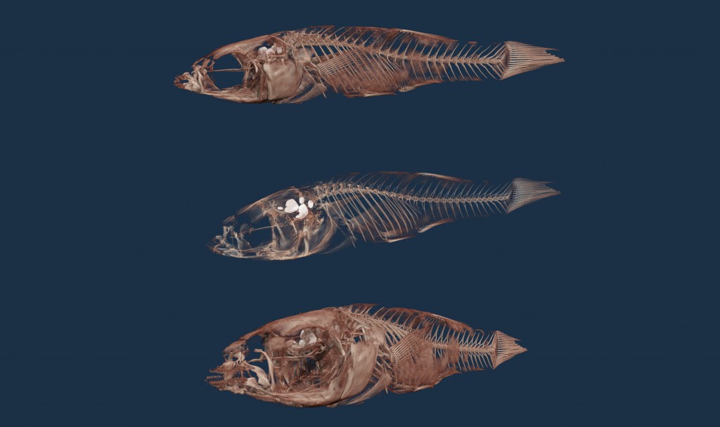
Micro computed tomography (CT) is a 3D X-ray imaging method similar to clinical CT scanners, but on a smaller scale with massively increased resolution. It enables 3D microscopy, in which every fine internal structure of an animal can be imaged non-destructively.
A micro-focus X-ray source illuminates the object and a planar X-ray detector collects hundreds of magnified projection images. A virtual 3D model is then reconstructed, simple or complex volumes of interest selected, and 3D morphometric parameters measured.
The Bruker Skyscan 1176 is a high performance in vivo micro-CT scanner for preclinical research. Its large 11 Megapixel X-ray camera delivers an unrivalled combination of resolution, image field size and scan speed.
It offers scanning of small animals, up to full rat body, at pixel sizes of 9, 18 and 35µm. Variable X-ray voltage and filters provide the flexibility to observe samples from soft tissue (lung) to bone/skeleton with metal implants.

