This webinar was recorded on May 20, 2015.
Seminar Overview
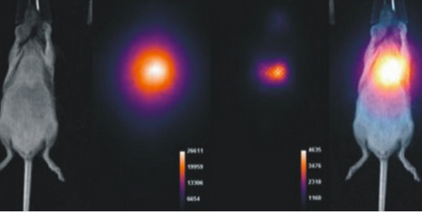
Sensitive in vivo luminescent imaging systems like the Bruker In-Vivo Xtreme may be leveraged not only for traditional bioluminescent imaging applications, but also for detection of PET radionuclide Cherenkov radiation. Cherenkov luminescent imaging (CLI) solutions, relative to traditional PET detector technology, provide an inexpensive approach to PET tracer imaging. Some have speculated that CLI technologies could supply broader access for more researchers to perform PET tracer studies. Still, CLI is limited in sensitivity (CL blue light emissions are subject to significant scatter in tissue) and is not applicable for some of the most common radioisotopes used in nuclear medicine (e.g. 99mTc).
The Bruker In-Vivo Xtreme system provides for multimodal X-ray imaging, fluorescence imaging (FLI), bioluminescence/Cherenkov imaging (BLI/CLI), and direct radioisotopic imaging (DRI), and is the only preclinical system offering combined CLI and DRI. The In-Vivo Xtreme performs DRI using a phosphor conversion screen for direct detection of radionuclide gamma ray emissions and overcomes many of the limitations of CLI, allowing for more sensitive detections and imaging of a broader range of isotopes.
What you will discover
During this webinar we will overview the multimodal capabilities of the In-Vivo Xtreme, highlighting the application potential for DRI.
Who should attend
Researchers involved in preclinical nuclear medicine, small-molecular compound studies, and in-vivo imaging.
Presenter
Todd Sasser,
PhD
Field Applications Scientist
Bruker Preclinical Imaging

