“In a new application note available from Bruker, Anna Mechling, Jürgen Hennig, Dominik von Elverfeldt and Laura Adela Harsan of the Department of Radiology, Medical Physics, University Hospital Freiburg, Germany discuss the findings of recent research into resting state functional MRI.”
Resting state functional MRI (rs-fMRI) is a method of functional brain imaging that can be used to evaluate regional interactions that occur when a subject is not performing a specific task. These interactions are observed through changes in the blood flow in the brain, creating a blood oxygen level dependent (BOLD) signal. Rs-MRI detects low frequency fluctuations of BOLD signal and their temporal correlations. The technique provides a non-invasive window into the intrinsic whole brain functional connectivity architecture, moving away from localizing isolated functional brain areas.
Rs-MRI is used extensively in human brain investigations and while the characterization of consistent and robust resting state networks in rodents is progressively evolving, fine-grained mapping of the mouse brain resting state networks remains an underexplored area. This is in contrast to the widespread use of this specie in experimental neuroscience for modeling human neurological disorders. Therefore, uncovering information related to the remodeling of brain networks in these models could contribute to a change in the view on brain disorders pathophysiology and finally establishing imaging biomarkers. A major challenge of rs-fMRI in small animals is the difficulty of achieving fast acquisition methods at high resolution while ensuring stable physiological parameters throughout the experiment. In this study, we demonstrate use of the CryoProbe, the latest technology for mouse MRI that provides a signal to noise increase of >2. This is achieved by the cooling of the probe and its electronics, making it possible to gain a fine-grained, non-invasive insight into the mouse brain functional “connectome”.

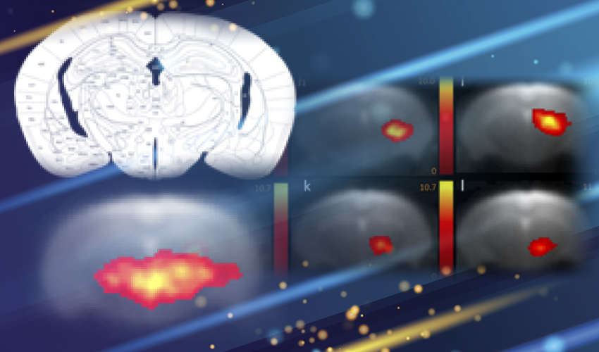
BOLD resting-state fMRI based cortical (A) and thalamic (B) mouse brain functional connectivity clusters, comparatively revealed by 40- (a, f) and 100-ICASSO (b–e and g–l). All images represent spatial color-coded z-maps of the independent components.
Based on this data, reliable patterns of the mouse brain intrinsic brain functional network architecture were identified. Our approach relies on the blind source spatial separation of the BOLD signal via group level independent component analysis (ICA) combined with it with graph theoretical analysis. We demonstrate that under medetomidine light sedation (adapted from Pawela et al., 2009) the basic modular properties known from the human brain were reproduced in the mouse and the network has the hallmarks of a small-world efficient network. We additionally distinguish the highly connected mouse brain areas, highlighting their importance as relays of the functional connectivity. This type of information is essential in translational studies, bridging the gap between preclinical and clinical investigations of functional connectome remodeling.
