Toward richer in vivo imaging data – a sequential protocol for trimodal PET/CT/SPECT imaging
Combining in vivo nuclear imaging with X-ray computed tomography (CT) in the same specimens provides a multi-dimensioned view into associated molecular, functional, and anatomical changes. To further enrich preclinical imaging studies, researchers have begun imaging dual PET or SPECT probes to track two related disease or pharmacologic targets within study cohorts. However, the combined use of PET and SPECT tracers together in the same animals has proven problematic due to cross-talk of the PET emissions generating noise in the SPECT results. In a recent paper, Chapman et al. present a protocol for combined PET and SPECT scans that overcomes this issue using sequential dosing and acquisition on a trimodal imaging system.
To investigate PET and SPECT probe/acquisition interactions, Chapman et al. first tested 96-well phantoms containing varying dilutions of probes containing the common SPECT and PET isotopes 99mTc and 18F. The results confirmed that addition the PET probe introduced artifacts into SPECT acquisitions, while the converse did not create noise. The authors propose that the PET/18F gamma rays penetrated the collimator and down-scattered into the SPECT camera, causing spurious detection events. In contrast, the PET acquisitions correctly excluded any non-paired events coming from the SPECT probe.
To prevent this tracer cross-talk in vivo, Chapman and colleagues devised a sequential scheme using a trimodal SPECT/CT/PET imaging system. The protocol started with SPECT dosing and acquisition, followed by CT, then PET dosing and lastly, imaging, so that no PET probe was present to interfere with SPECT imaging. By capturing all three modalities on the same trimodal platform, the researchers avoided moving the animals, enabling high-accuracy, automated co-registration of the PET, SPECT, and CT results.
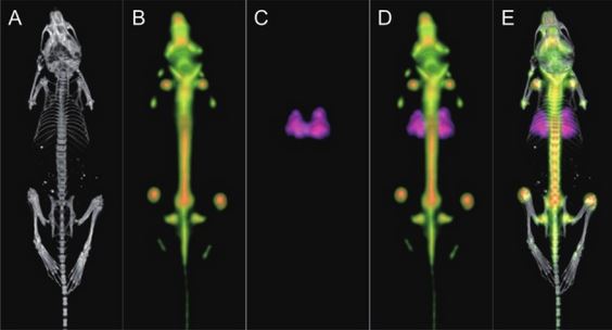
The research team used commercially available SPECT and PET tracers that accumulate at sites of lung perfusion and bone regeneration, respectively. Using the Bruker Albira trimodal PET/SPECT/CT imaging system, the team first dosed male hairless mice with SPECT tracer (99mTc -MAA or 99mTc -Pentetate) via a tail vein catheter. After 30 minutes for probe biodistribution, the researchers performed a 10-minute SPECT acquisition (120 mm FOV, multi-pinhole collimator, 60 projections), followed by CT (110 mm FOV, 45 kVp, 200 uA, at 400 projections). Next, the team gave a dose of PET tracer (Na18F), allowing 30 minutes for distribution before capturing a PET image with a five-minute integration time (110 mm FOV). The team used Albira Suite Reconstructor software to automatically fuse the SPECT and CT data, then the CT and PET images.
The final, fused images show clear lung and bone labeling aligned over the CT image, clear of PET/SPECT cross-talk. The authors emphasize that the trimodal imaging system made co-registration of PET, SPECT, and CT data straightforward by preventing the need to move animals between modalities.
This widely applicable method can help save time and enhance information gained in preclinical in vivo studies by supporting concurrent exploration of related physiological and molecular functions – such as perfusion, neurotransmission, metabolism, apoptosis, angiogenesis – under identical conditions. Using commercially available PET and SPECT tracers and the Albira SPECT/CT/PET system, this new protocol may be particularly useful for applications in neurology, oncology, and cardiac imaging.
Reference
Chapman SE, Diener JM, Sasser TA, Correcher C, González AJ, Van Avermaete T, Leevy WM. Dual tracer imaging of SPECT and PET probes in living mice using a sequential protocol. Am J Nucl Med Mol Imaging. 2012;2:405–413. PubMed Central: PMC3484419.
